Search Completed | Title | PEMF Trigger Cell Death and Senescence in Cancer Cells
Original File Name Searched: ijms-25-02473-v2.pdf | Google It | Yahoo | Bing
Page | 007 Int. J. Mol. Sci. 2024, 25, 2473
7 of 14
the low-intensity, frequency-modulated (6–25 Hz) Thomas electromagnetic field (EMF)
pattern [21]. The Thomas EMF was able to inhibit the growth of cancer cell lines including
B16-BL6, MCF-7, MDA-MB-231 and HeLa via increased Ca2+ uptake through T-type Ca2+
channels but did not affect the growth of normal cells. Additionally, Crocetti and colleagues
showed similar results in an in vitro PEMF treatment study using cancer and non-cancer
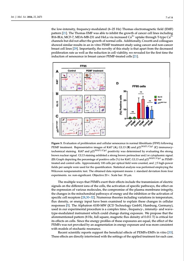
Figure 5. Evaluation of proliferation and cellular senescence in normal fibroblasts (FF95) following Figure 5. Evaluation of proliferation and cellular senescence in normal fibroblasts (FF95) following
PEMF treatment. Representative images of Ki67 (A), GL13 (B) and p21WAF1/Cip1 (C) immunocytochem- PEMF treatment. Representative images of Ki67 (A), GL13 (B) and p21WAF1/Cip1 (C) immunocy- ical staining. Ki67 and p21WAF1/Cip1 positivity was determined by evaluating the strong brown nuclear tochemical staining. Ki67 and p21WAF1/Cip1 positivity was determined by evaluating the strong signal. GL13 staining exhibited a strong brown perinuclear and/or cytoplasmic signal. (D) Graph
brownnuclearsignal.GL13stainingexhibitedastrongbrownperinucleWaArF1a/Cnipd1/orcytoplasmicsignal. depicting the percentage of positive cells (%) for Ki67, GL13 and p21 in PEMF-treated and
(D) Graph depicting the percentage of positive cells (%) for Ki67, GL13 and p21WAF1/Cip1 in PEMF- control cells. Approximately 100 cells per optical field were counted, and ≥5 high-power fields per
breast cell lines [29]. Importantly, the novelty of this study is that apart from the decreased Int. J. Mol. Sci. 2024, 25, x FOR PEER REVIEW 8 of 15
proliferation rate as well as the reduction in cell viability, we revealed for the first time the induction of senescence in breast cancer PEMF-treated cells [21].
trseaamtepdleawnderceounstreodlfcoerlltsh.eAqpuparnotxifiimcattieolny.1S0ta0tcisetlilcsaplearnaolpytsiicsawl fiaeslpdewrfeoremceodunemtepdl,oaynidng≥t5hehWighil-cpooxwoner nonparametric test. The obtained data represent means ± standard deviation from four experiments.
fields per sample were used for the quantification. Statistical analysis was performed employing the
ns: non-significant. Objective 20×. Scale bar: 30 μm.
Wilcoxon nonparametric test. The obtained data represent means ± standard deviation from four experiments. ns: non-significant. Objective 20×. Scale bar: 30 μm.
The multiple ways that PEMFs exert their effects include the transmission of electric signals on the different ions of the cells, the activation of specific pathways, the effect on
The multiple ways that PEMFs exert their effects include the transmission of electric
the expression of various molecules, the compromise of the plasma membrane integrity,
signals on the different ions of the cells, the activation of specific pathways, the effect on
the changes in the mitochondrial pathways of energy and the inhibition or the activation
the expression of various molecules, the compromise of the plasma membrane integrity,
of specific cell receptors [28–31]. Numerous theories including variations in temperature,
the changes in the mitochondrial pathways of energy and the inhibition or the activation of
flux density, or energy input have been examined to explain these changes in cellular re-
specific cell receptors [28,30–32]. Numerous theories including variations in temperature, sponses [5]. The Alphatron 4100-MW (JCD Technology GmbH, Hamburg, Germany),
flux density, or energy input have been examined to explain these changes in cellular
used in our experimental procedure is a complex time-, frequency-, intensity- and wave-
responses [5]. The Alphatron 4100-MW (JCD Technology GmbH, Hamburg, Germany), type-modulated instrument which could change during exposure. We propose that the
used in our experimental procedure is a complex time-, frequency-, intensity- and wave-
aforementioned pattern (8 Hz, full square, magnetic flux density of 0.011 T) is critical for
type-modulated instrument which could change during exposure. We propose that the
its effects on cells. Since the energy profiles of these exposures are equal, the effect of the
aforementioned pattern (8 Hz, full square, magnetic flux density of 0.011 T) is critical for
PEMFs was not provoked by an augmentation in energy exposure and was more con-
its effects on cells. Since the energy profiles of these exposures are equal, the effect of the
sistent with models of stochastic resonance.
PEMFs was not provoked by an augmentation in energy exposure and was more consistent
Recent scientific reports support the beneficial effects of PEMFs-EMFs in vitro [32].
with models of stochastic resonance.
These effects are directly intertwined with the settings of the applied treatment for each
Recent scientific reports support the beneficial effects of PEMFs-EMFs in vitro [33]. case. Specifically, the beneficial biological effects that have been reported are partially as-
These effects are directly intertwined with the settings of the applied treatment for each case.
cribed not only to the amplitude, frequency, modulation, intensity, wave type and dura- tion of exposure of the applied PEMF-EMF program, but also to the biological systems [33,34]. However, the biological mechanisms that have been proposed to explain the ac- tion of PEMFs-EMFs in biological systems are restricted [35,36]. In the work of Ayşe et al., an ELF-PEMF program of 50 Hz and 100 G exposure [37] induced the apoptosis and ne- crosis of K562 and U937 cells; noteworthily, a proliferation decrease (15% approximately)
|