PDF Publication Title:
Text from PDF Page: 114
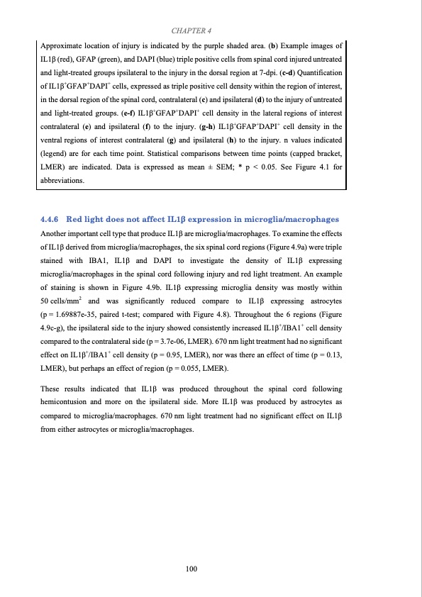
CHAPTER 4 Approximate location of injury is indicated by the purple shaded area. (b) Example images of IL1β (red), GFAP (green), and DAPI (blue) triple positive cells from spinal cord injured untreated and light-treated groups ipsilateral to the injury in the dorsal region at 7-dpi. (c-d) Quantification of IL1β+GFAP+DAPI+ cells, expressed as triple positive cell density within the region of interest, in the dorsal region of the spinal cord, contralateral (c) and ipsilateral (d) to the injury of untreated and light-treated groups. (e-f) IL1β+GFAP+DAPI+ cell density in the lateral regions of interest contralateral (e) and ipsilateral (f) to the injury. (g-h) IL1β+GFAP+DAPI+ cell density in the ventral regions of interest contralateral (g) and ipsilateral (h) to the injury. n values indicated (legend) are for each time point. Statistical comparisons between time points (capped bracket, LMER) are indicated. Data is expressed as mean ± SEM; * p < 0.05. See Figure 4.1 for abbreviations. 4.4.6 Red light does not affect IL1β expression in microglia/macrophages Another important cell type that produce IL1β are microglia/macrophages. To examine the effects of IL1β derived from microglia/macrophages, the six spinal cord regions (Figure 4.9a) were triple stained with IBA1, IL1β and DAPI to investigate the density of IL1β expressing microglia/macrophages in the spinal cord following injury and red light treatment. An example of staining is shown in Figure 4.9b. IL1β expressing microglia density was mostly within 50cells/mm2 and was significantly reduced compare to IL1β expressing astrocytes (p = 1.69887e-35, paired t-test; compared with Figure 4.8). Throughout the 6 regions (Figure 4.9c-g), the ipsilateral side to the injury showed consistently increased IL1β+/IBA1+ cell density compared to the contralateral side (p = 3.7e-06, LMER). 670 nm light treatment had no significant effect on IL1β+/IBA1+ cell density (p = 0.95, LMER), nor was there an effect of time (p = 0.13, LMER), but perhaps an effect of region (p = 0.055, LMER). These results indicated that IL1β was produced throughout the spinal cord following hemicontusion and more on the ipsilateral side. More IL1β was produced by astrocytes as compared to microglia/macrophages. 670 nm light treatment had no significant effect on IL1β from either astrocytes or microglia/macrophages. 100PDF Image | Effects of Red Light Treatment on Spinal Cord Injury
PDF Search Title:
Effects of Red Light Treatment on Spinal Cord InjuryOriginal File Name Searched:
Thesis_Di Hu_final.pdfDIY PDF Search: Google It | Yahoo | Bing
Cruise Ship Reviews | Luxury Resort | Jet | Yacht | and Travel Tech More Info
Cruising Review Topics and Articles More Info
Software based on Filemaker for the travel industry More Info
The Burgenstock Resort: Reviews on CruisingReview website... More Info
Resort Reviews: World Class resorts... More Info
The Riffelalp Resort: Reviews on CruisingReview website... More Info
| CONTACT TEL: 608-238-6001 Email: greg@cruisingreview.com | RSS | AMP |