PDF Publication Title:
Text from PDF Page: 003
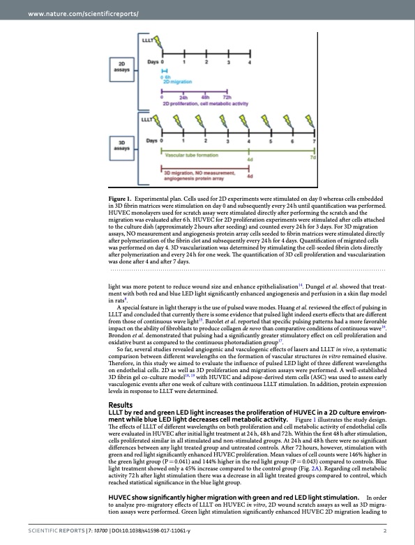
www.nature.com/scientificreports/ Figure 1. Experimental plan. Cells used for 2D experiments were stimulated on day 0 whereas cells embedded in 3D fibrin matrices were stimulation on day 0 and subsequently every 24 h until quantification was performed. HUVEC monolayers used for scratch assay were stimulated directly after performing the scratch and the migration was evaluated after 6 h. HUVEC for 2D proliferation experiments were stimulated after cells attached to the culture dish (approximately 2 hours after seeding) and counted every 24 h for 3 days. For 3D migration assays, NO measurement and angiogenesis protein array cells seeded to fibrin matrices were stimulated directly after polymerization of the fibrin clot and subsequently every 24 h for 4 days. Quantification of migrated cells was performed on day 4. 3D vascularization was determined by stimulating the cell-seeded fibrin clots directly after polymerization and every 24 h for one week. The quantification of 3D cell proliferation and vascularization was done after 4 and after 7 days. light was more potent to reduce wound size and enhance epithelialisation14. Dungel et al. showed that treat- ment with both red and blue LED light significantly enhanced angiogenesis and perfusion in a skin flap model in rats8. A special feature in light therapy is the use of pulsed wave modes. Huang et al. reviewed the effect of pulsing in LLLT and concluded that currently there is some evidence that pulsed light indeed exerts effects that are different from those of continuous wave light15. Barolet et al. reported that specific pulsing patterns had a more favorable impact on the ability of fibroblasts to produce collagen de novo than comparative conditions of continuous wave16. Brondon et al. demonstrated that pulsing had a significantly greater stimulatory effect on cell proliferation and oxidative burst as compared to the continuous photoradiation group17. So far, several studies revealed angiogenic and vasculogenic effects of lasers and LLLT in vivo, a systematic comparison between different wavelengths on the formation of vascular structures in vitro remained elusive. Therefore, in this study we aimed to evaluate the influence of pulsed LED light of three different wavelengths on endothelial cells. 2D as well as 3D proliferation and migration assays were performed. A well-established 3D fibrin gel co-culture model18, 19 with HUVEC and adipose-derived stem cells (ASC) was used to assess early vasculogenic events after one week of culture with continuous LLLT stimulation. In addition, protein expression levels in response to LLLT were determined. Results LLLT by red and green LED light increases the proliferation of HUVEC in a 2D culture environ- ment while blue LED light decreases cell metabolic activity. Figure 1 illustrates the study design. The effects of LLLT of different wavelengths on both proliferation and cell metabolic activity of endothelial cells were evaluated in HUVEC after initial light treatment at 24 h, 48 h and 72 h. Within the first 48 h after stimulation, cells proliferated similar in all stimulated and non-stimulated groups. At 24 h and 48 h there were no significant differences between any light treated group and untreated controls. After 72 hours, however, stimulation with green and red light significantly enhanced HUVEC proliferation. Mean values of cell counts were 146% higher in the green light group (P = 0.041) and 144% higher in the red light group (P = 0.043) compared to controls. Blue light treatment showed only a 45% increase compared to the control group (Fig. 2A). Regarding cell metabolic activity 72 h after light stimulation there was a decrease in all light treated groups compared to control, which reached statistical significance in the blue light group. HUVEC show significantly higher migration with green and red LED light stimulation. In order to analyze pro-migratory effects of LLLT on HUVEC in vitro, 2D wound scratch assays as well as 3D migra- tion assays were performed. Green light stimulation significantly enhanced HUVEC 2D migration leading to SCientifiC REpORTS | 7: 10700 | DOI:10.1038/s41598-017-11061-y 2PDF Image | impact of wavelengths of LED light-therapy on endothelial cells
PDF Search Title:
impact of wavelengths of LED light-therapy on endothelial cellsOriginal File Name Searched:
Rohringeretal-LLLT2017.pdfDIY PDF Search: Google It | Yahoo | Bing
Cruise Ship Reviews | Luxury Resort | Jet | Yacht | and Travel Tech More Info
Cruising Review Topics and Articles More Info
Software based on Filemaker for the travel industry More Info
The Burgenstock Resort: Reviews on CruisingReview website... More Info
Resort Reviews: World Class resorts... More Info
The Riffelalp Resort: Reviews on CruisingReview website... More Info
| CONTACT TEL: 608-238-6001 Email: greg@cruisingreview.com | RSS | AMP |