PDF Publication Title:
Text from PDF Page: 011
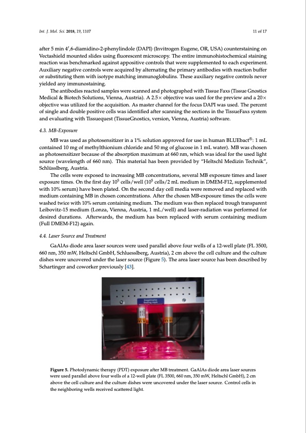
Int. J. Mol. Sci. 2018, 19, 1107 11 of 17 after 5 min 4′,6-diamidino-2-phenylindole (DAPI) (Invitrogen Eugene, OR, USA) counterstaining on Vectashield mounted slides using fluorescent microscopy. The entire immunohistochemical staining reaction was benchmarked against appositive controls that were supplemented to each experiment. Auxiliary negative controls were acquired by alternating the primary antibodies with reaction buffer or substituting them with isotype matching immunoglobulins. These auxiliary negative controls never yielded any immunostaining. The antibodies reacted samples were scanned and photographed with Tissue Faxs (Tissue Gnostics Medical & Biotech Solutions, Vienna, Austria). A 2.5× objective was used for the preview and a 20× objective was utilized for the acquisition. As master channel for the focus DAPI was used. The percent of single and double positive cells was identified after scanning the sections in the TissueFaxs system and evaluating with Tissuequest (TissueGnostics, version, Vienna, Austria) software. 4.3. MB-Exposure MB was used as photosensitizer in a 1% solution approved for use in human BLUEbact®: 1 mL contained 10 mg of methylthionium chloride and 50 mg of glucose in 1 mL water). MB was chosen as photosensitizer because of the absorption maximum at 660 nm, which was ideal for the used light source (wavelength of 660 nm). This material has been provided by “Heltschl Medizin Technik”, Schlüsslberg, Austria. The cells were exposed to increasing MB concentrations, several MB exposure times and laser exposure times. On the first day 105 cells/well (105 cells/2 mL medium in DMEM-F12, supplemented with 10% serum) have been plated. On the second day cell media were removed and replaced with medium containing MB in chosen concentrations. After the chosen MB-exposure times the cells were washed twice with 10% serum containing medium. The medium was then replaced trough transparent Leibovitz-15 medium (Lonza, Vienna, Austria, 1 mL/well) and laser-radiation was performed for desired durations. Afterwards, the medium has been replaced with serum containing medium (Full DMEM-F12) again. 4.4. Laser Source and Treatment GaAlAs diode area laser sources were used parallel above four wells of a 12-well plate (FL 3500, 660 nm, 350 mW, Heltschl GmbH, Schluesslberg, Austria), 2 cm above the cell culture and the culture dishes were uncovered under the laser source (Figure 5). The area laser source has been described by Schartinger and coworker previously [43]. Figure 5. Photodynamic therapy (PDT) exposure after MB treatment. GaAlAs diode area laser sources were used parallel above four wells of a 12-well plate (FL 3500, 660 nm, 350 mW, Heltschl GmbH), 2 cm above the cell culture and the culture dishes were uncovered under the laser source. Control cells in the neighboring wells received scattered light.PDF Image | Photodynamic Low Level Laser Squamous Cell Carcinoma
PDF Search Title:
Photodynamic Low Level Laser Squamous Cell CarcinomaOriginal File Name Searched:
ijms-19-01107.pdfDIY PDF Search: Google It | Yahoo | Bing
Cruise Ship Reviews | Luxury Resort | Jet | Yacht | and Travel Tech More Info
Cruising Review Topics and Articles More Info
Software based on Filemaker for the travel industry More Info
The Burgenstock Resort: Reviews on CruisingReview website... More Info
Resort Reviews: World Class resorts... More Info
The Riffelalp Resort: Reviews on CruisingReview website... More Info
| CONTACT TEL: 608-238-6001 Email: greg@cruisingreview.com | RSS | AMP |