PDF Publication Title:
Text from PDF Page: 003
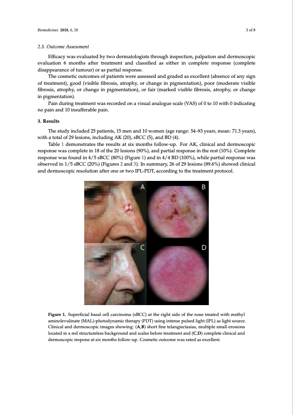
Biomedicines 2018, 6, 18 3 of 8 Biomedicines 2018, 6, x 3 of 8 2.3. Outcome Assessment Effificacy was evaluated by two dermatologists through inspection, palpation and dermoscopic evaluation 6 months after treatment and classiffiied as either in complete response (complete disappearance of tumour) or as partial response. The cosmetic outcomes of patients were assessed and graded as excellent (absence of any sign of treatment), good (visible fifibrosis, atrophy, or change in piigmentation), poor (moderate visible fifibrosis,atrophy,orchangeiinppigigmeenntatatitoionn),),ororfafiarir(m(marakrekdedvivsibsilbelfeibfirborsois,isa,traotprohpyh,yo,rocrhacnhganegine ipnigpmigemnteanttiaotnio).n). Paindurrininggtrteraetamtmenetnwtawsarsecroercdoerdeodnaonvisauavliasunaloagnuaeloscgaule(sVcAalSe)o(Vf0AtSo)1o0fw0ithto01in0dwicaitthing0 ninodpicaaitninagndno10paininsuafnfedra1b0leinpsuaifnfe. rable pain. 3. Results 3. Results The study included 25 patients, 15 men and 10 women (age range: 54–93 years, mean: 71.3 years), The study included 25 patients, 15 men and 10 women (age range: 54–93 years, mean: 71.3 with a total of 29 lesions, including AK (20), sBCC (5), and BD (4). years), with a total of 29 lesions, including AK (20), sBCC (5), and BD (4). Table 1 demonstrates the results at six months follow-up. For AK, clinical and dermoscopic Table 1 demonstrates the results at six months follow-up. For AK, clinical and dermoscopic response was complete in 18 of the 20 lesions (90%), and partial response in the rest (10%). Complete response was complete in 18 of the 20 lesions (90%), and partial response in the rest (10%). Complete response was found in 4/5 sBCC (80%) (Figure 1) and in 4/4 BD (100%), while partial response was response was found in 4/5 sBCC (80%) (Figure 1) and in 4/4 BD (100%), while partial response was observed in 1/5 sBCC (20%) (Figures 2 and 3). In summary, 26 of 29 lesions (89.6%) showed clinical observed in 1/5 sBCC (20%) (Figures 2 and 3). In summary, 26 of 29 lesions (89.6%) showed clinical and dermoscopic resolution after one or two IPL-PDT, according to the treatment protocol. and dermoscopic resolution after one or two IPL-PDT, according to the treatment protocol. Figure 1. Superficial basal cell carcinoma (sBCC) at the right side of the nose treated with methyl Figure 1. Superficial basal cell carcinoma (sBCC) at the right side of the nose treated with methyl aminolevulinate (MAL)-photodynamic therapy (PDT) using intense pulsed light (IPL) as light aminolevulinate (MAL)-photodynamic therapy (PDT) using intense pulsed light (IPL) as light source. source. Clinical and dermoscopic images showing: (A,B) short fine telangiectasias, multiple small Clinical and dermoscopic images showing: (A,B) short fine telangiectasias, multiple small erosions erosions located in a red structureless background and scales before treatment and (C,D) complete located in a red structureless background and scales before treatment and (C,D) complete clinical and clinical and dermoscopic respone at six months follow-up. Cosmetic outcome was rated as excellent. dermoscopic respone at six months follow-up. Cosmetic outcome was rated as excellent.PDF Image | Photodynamic Therapy Pulsed Light Treatment of Nonmelanoma
PDF Search Title:
Photodynamic Therapy Pulsed Light Treatment of NonmelanomaOriginal File Name Searched:
biomedicines-06-00018.pdfDIY PDF Search: Google It | Yahoo | Bing
Cruise Ship Reviews | Luxury Resort | Jet | Yacht | and Travel Tech More Info
Cruising Review Topics and Articles More Info
Software based on Filemaker for the travel industry More Info
The Burgenstock Resort: Reviews on CruisingReview website... More Info
Resort Reviews: World Class resorts... More Info
The Riffelalp Resort: Reviews on CruisingReview website... More Info
| CONTACT TEL: 608-238-6001 Email: greg@cruisingreview.com | RSS | AMP |