PDF Publication Title:
Text from PDF Page: 003
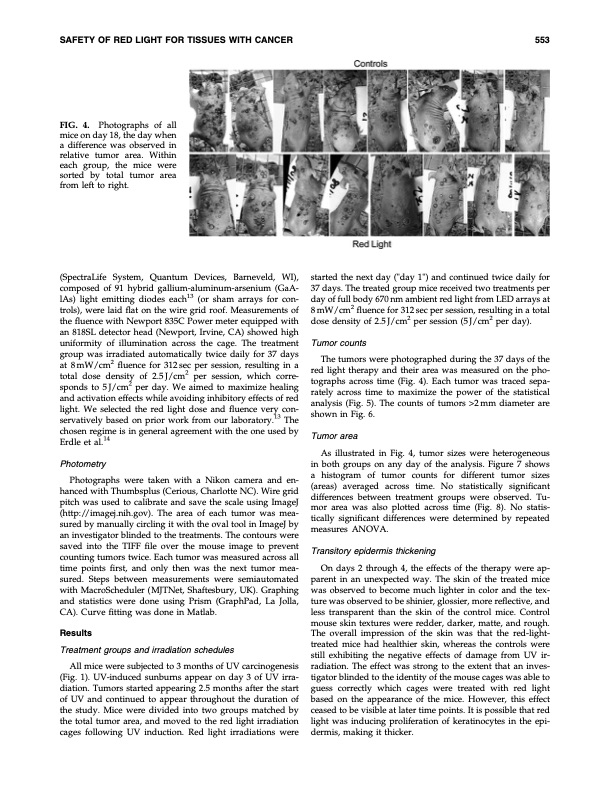
SAFETY OF RED LIGHT FOR TISSUES WITH CANCER 553 FIG. 4. Photographs of all mice on day 18, the day when a difference was observed in relative tumor area. Within each group, the mice were sorted by total tumor area from left to right. (SpectraLife System, Quantum Devices, Barneveld, WI), composed of 91 hybrid gallium-aluminum-arsenium (GaA- lAs) light emitting diodes each13 (or sham arrays for con- trols), were laid flat on the wire grid roof. Measurements of the fluence with Newport 835C Power meter equipped with an 818SL detector head (Newport, Irvine, CA) showed high uniformity of illumination across the cage. The treatment group was irradiated automatically twice daily for 37 days at 8mW/cm2 fluence for 312sec per session, resulting in a total dose density of 2.5J/cm2 per session, which corre- sponds to 5 J/cm2 per day. We aimed to maximize healing and activation effects while avoiding inhibitory effects of red light. We selected the red light dose and fluence very con- servatively based on prior work from our laboratory.13 The chosen regime is in general agreement with the one used by Erdle et al.14 Photometry Photographs were taken with a Nikon camera and en- hanced with Thumbsplus (Cerious, Charlotte NC). Wire grid pitch was used to calibrate and save the scale using ImageJ (http://imagej.nih.gov). The area of each tumor was mea- sured by manually circling it with the oval tool in ImageJ by an investigator blinded to the treatments. The contours were saved into the TIFF file over the mouse image to prevent counting tumors twice. Each tumor was measured across all time points first, and only then was the next tumor mea- sured. Steps between measurements were semiautomated with MacroScheduler ( MJTNet, Shaftesbury, UK). Graphing and statistics were done using Prism (GraphPad, La Jolla, CA). Curve fitting was done in Matlab. Results Treatment groups and irradiation schedules All mice were subjected to 3 months of UV carcinogenesis (Fig. 1). UV-induced sunburns appear on day 3 of UV irra- diation. Tumors started appearing 2.5 months after the start of UV and continued to appear throughout the duration of the study. Mice were divided into two groups matched by the total tumor area, and moved to the red light irradiation cages following UV induction. Red light irradiations were started the next day ("day 1") and continued twice daily for 37 days. The treated group mice received two treatments per day of full body 670 nm ambient red light from LED arrays at 8 mW/cm2 fluence for 312 sec per session, resulting in a total dose density of 2.5 J/cm2 per session (5 J/cm2 per day). Tumor counts The tumors were photographed during the 37 days of the red light therapy and their area was measured on the pho- tographs across time (Fig. 4). Each tumor was traced sepa- rately across time to maximize the power of the statistical analysis (Fig. 5). The counts of tumors >2mm diameter are shown in Fig. 6. Tumor area As illustrated in Fig. 4, tumor sizes were heterogeneous in both groups on any day of the analysis. Figure 7 shows a histogram of tumor counts for different tumor sizes (areas) averaged across time. No statistically significant differences between treatment groups were observed. Tu- mor area was also plotted across time (Fig. 8). No statis- tically significant differences were determined by repeated measures ANOVA. Transitory epidermis thickening On days 2 through 4, the effects of the therapy were ap- parent in an unexpected way. The skin of the treated mice was observed to become much lighter in color and the tex- ture was observed to be shinier, glossier, more reflective, and less transparent than the skin of the control mice. Control mouse skin textures were redder, darker, matte, and rough. The overall impression of the skin was that the red-light- treated mice had healthier skin, whereas the controls were still exhibiting the negative effects of damage from UV ir- radiation. The effect was strong to the extent that an inves- tigator blinded to the identity of the mouse cages was able to guess correctly which cages were treated with red light based on the appearance of the mice. However, this effect ceased to be visible at later time points. It is possible that red light was inducing proliferation of keratinocytes in the epi- dermis, making it thicker.PDF Image | Preliminary Study of the Safety of Red Light Phototherapy Cancer
PDF Search Title:
Preliminary Study of the Safety of Red Light Phototherapy CancerOriginal File Name Searched:
phototherapy-for-cancer.pdfDIY PDF Search: Google It | Yahoo | Bing
Cruise Ship Reviews | Luxury Resort | Jet | Yacht | and Travel Tech More Info
Cruising Review Topics and Articles More Info
Software based on Filemaker for the travel industry More Info
The Burgenstock Resort: Reviews on CruisingReview website... More Info
Resort Reviews: World Class resorts... More Info
The Riffelalp Resort: Reviews on CruisingReview website... More Info
| CONTACT TEL: 608-238-6001 Email: greg@cruisingreview.com | RSS | AMP |