PDF Publication Title:
Text from PDF Page: 004
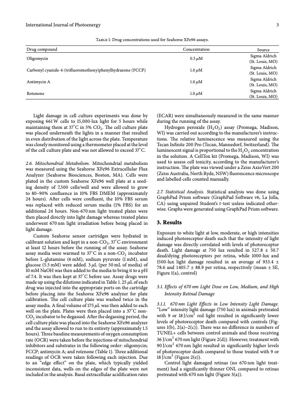
International Journal of Photoenergy 3 Drug compound Oligomycin Carbonyl cyanide-4-(trifluoromethoxy)phenylhydrazone (FCCP) Antimycin A Rotenone Light damage in cell culture experiments was done by exposing 661 W cells to 15,000-lux light for 5 hours while maintaining them at 37∘ C in 5% CO2 . The cell culture plate was placed underneath the lights in a manner that resulted in even distribution of the light across the plate. Temperature was closely monitored using a thermometer placed at the level of the cell culture plate and was not allowed to exceed 37∘C. 2.6. Mitochondrial Metabolism. Mitochondrial metabolism was measured using the Seahorse XFe96 Extracellular Flux Analyzer (Seahorse Biosciences, Boston, MA). Cells were plated in the custom Seahorse XFe96 well plate at a seed- ing density of 7,500 cells/well and were allowed to grow to 80–90% confluence in 10% FBS DMEM (approximately 24 hours). After cells were confluent, the 10% FBS serum was replaced with reduced serum media (1% FBS) for an additional 24 hours. Non-670 nm light treated plates were then placed directly into light damage whereas treated plates underwent 670 nm light irradiation before being placed in light damage. Custom Seahorse sensor cartridges were hydrated in calibrant solution and kept in a non-CO2, 37∘C environment at least 12 hours before the running of the assay. Seahorse assay media were warmed to 37∘C in a non-CO2 incubator before L-glutamine (6mM), sodium pyruvate (1mM), and glucose (5.5 mM) were added. 3 𝜇L (per 50 mL of media) of 10 mM NaOH was then added to the media to bring it to a pH of 7.4. It was then kept at 37∘C before use. Assay drugs were made up using the dilutions indicated in Table 1. 25 𝜇L of each drug was injected into the appropriate ports on the cartridge before placing into the Seahorse XFe96 analyzer for plate calibration. The cell culture plate was washed twice in the assay media. A final volume of 175 𝜇L was then added to each well on the plate. Plates were then placed into a 37∘C non- CO2 incubator to be degassed. After the degassing period, the cell culture plate was placed into the Seahorse XFe96 analyzer and the assay allowed to run to its entirety (approximately 1.5 hours). Three baseline measurements of oxygen consumption rate (OCR) were taken before the injections of mitochondrial inhibitors and substrates in the following order: oligomycin; FCCP; antimycin A; and rotenone (Table 1). Three additional readings of OCR were taken following each injection. Due to an “edge effect” on the plate, which typically yielded inconsistent data, wells on the edges of the plate were not included in the analysis. Basal extracellular acidification rates Concentration 0.5 𝜇M 1.0 𝜇M 1.0 𝜇M 1.0 𝜇M Source Sigma Aldrich (St. Louis, MO) Sigma Aldrich (St. Louis, MO) Sigma Aldrich (St. Louis, MO) Sigma Aldrich (St. Louis, MO) Table 1: Drug concentrations used for Seahorse XFe96 assays. (ECAR) were simultaneously measured in the same manner during the running of the assay. Hydrogen peroxide (H2O2) assay (Promega, Madison, WI) was carried out according to the manufacturer’s instruc- tions. The relative luminescence was measured using the Tecan Infinite 200 Pro (Tecan, Mannedorf, Switzerland). The luminescent signal is proportional to the H2O2 concentration in the solution. A CellTox kit (Promega, Madison, WI) was used to assess cell toxicity, according to the manufacturer’s instruction. The plate was viewed under a Zeiss AxioVert 200 (Zeiss Australia, North Ryde, NSW) fluorescence microscope and labelled cells counted manually. 2.7. Statistical Analysis. Statistical analysis was done using GraphPad Prism software (GraphPad Software v6, La Jolla, CA) using unpaired Student’s 𝑡-test unless indicated other- wise. Graphs were generated using GraphPad Prism software. 3. Results Exposure to white light at low, moderate, or high intensities induced photoreceptor death such that the intensity of light damage was directly correlated with levels of photoreceptor death. Light damage at 750 lux resulted in 527.8 ± 50.7 dead/dying photoreceptors per retina, while 1000-lux and 1500-lux light damage resulted in an average of 933.4 ± 78.6 and 1405.7 ± 88.9 per retina, respectively (mean ± SE, Figure 1(a), control) 3.1. Effects of 670 nm Light Dose on Low, Medium, and High Intensity Retinal Damage 3.1.1. 670 nm Light Effects in Low Intensity Light Damage. “Low” intensity light damage (750 lux) in animals pretreated with 9 or 18 J/cm2 red light resulted in significantly lower levels of photoreceptor death compared with controls (Fig- ures 1(b), 2(a)–2(c)). There was no difference in numbers of TUNEL+ cells between control animals and those receiving 36 J/cm2 670 nm light (Figure 2(d)). However, treatment with 90 J/cm2 670 nm light resulted in significantly higher levels of photoreceptor death compared to those treated with 9 or 18 J/cm2 (Figure 2(e)). Control light damaged retinas (no 670nm light treat- ment) had a significantly thinner ONL compared to retinas pretreated with 670 nm light (Figure 3(a)).PDF Image | 670 nm Light Therapy to Protect vs Photoreceptor Cell Death
PDF Search Title:
670 nm Light Therapy to Protect vs Photoreceptor Cell DeathOriginal File Name Searched:
Efficacy_of_670_nm_Light_Therapy_to_Protect_agains.pdfDIY PDF Search: Google It | Yahoo | Bing
Cruise Ship Reviews | Luxury Resort | Jet | Yacht | and Travel Tech More Info
Cruising Review Topics and Articles More Info
Software based on Filemaker for the travel industry More Info
The Burgenstock Resort: Reviews on CruisingReview website... More Info
Resort Reviews: World Class resorts... More Info
The Riffelalp Resort: Reviews on CruisingReview website... More Info
| CONTACT TEL: 608-238-6001 Email: greg@cruisingreview.com | RSS | AMP |