PDF Publication Title:
Text from PDF Page: 007
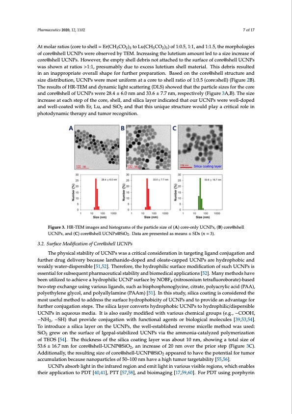
Pharmaceutics 2020, 12, 1102 7 of 17 At molar ratios (core to shell = Er(CH3CO2)3 to Lu(CH3CO2)3) of 1:0.5, 1:1, and 1:1.5, the morphologies Pharmaceutics 2020, 12, x FOR PEER REVIEW 7 of 16 of core@shell UCNPs were observed by TEM. Increasing the lutetium amount led to a size increase of core@shell UCNPs. However, the empty shell debris not attached to the surface of core@shell UCNPs core@shell UCNPs. However, the empty shell debris not attached to the surface of core@shell UCNPs was shown at ratios >1:1, presumably due to excess lutetium shell material. This debris resulted was shown at ratios >1:1, presumably due to excess lutetium shell material. This debris resulted in an in an inappropriate overall shape for further preparation. Based on the core@shell structure and inappropriate overall shape for further preparation. Based on the core@shell structure and size size distribution, UCNPs were most uniform at a core to shell ratio of 1:0.5 (core:shell) (Figure 2B). distribution, UCNPs were most uniform at a core to shell ratio of 1:0.5 (core:shell) (Figure 2B). The The results of HR-TEM and dynamic light scattering (DLS) showed that the particle sizes for the core results of HR-TEM and dynamic light scattering (DLS) showed that the particle sizes for the core and and core@shell of UCNPs were 28.4 ± 6.0 nm and 33.6 ± 7.7 nm, respectively (Figure 3A,B). The size core@shell of UCNPs were 28.4 ± 6.0 nm and 33.6 ± 7.7 nm, respectively (Figure 3A,B). The size increase at each step of the core, shell, and silica layer indicated that our UCNPs were well-doped increase at each step of the core, shell, and silica layer indicated that our UCNPs were well-doped and well-coated with Er, Lu, and SiO and that this unique structure would play a critical role in and well-coated with Er, Lu, and SiO2 and that this unique structure would play a critical role in photodynamic therapy and tumor recognition. photodynamic therapy and tumor recognition. Figure 3. HR-TEM images and histograms of the particle size of (A) core-only UCNPs, (B) core@shell UCNPs, and (C) core@shell UCNPs@SiO2..Daattaaaarreprresenttedasmeans±SSDss((nn==33).). 3.2. Surface Modification of Core@shell UCNPs 3.2. Surface Modification of Core@shell UCNPs 2 The physical stability of UCNPs was a critical consideration in targeting ligand conjugation and The physical stability of UCNPs was a critical consideration in targeting ligand conjugation and further drug delivery because lanthanide-doped and oleate-capped UCNPs are hydrophobic and further drug delivery because lanthanide-doped and oleate-capped UCNPs are hydrophobic and weakly water-dispersible [51,52]. Therefore, the hydrophilic surface modification of such UCNPs is weakly water-dispersible [51,52]. Therefore, the hydrophilic surface modification of such UCNPs is essential for subsequent pharmaceutical stability and biomedical applications [52]. Many methods have essential for subsequent pharmaceutical stability and biomedical applications [52]. Many methods been utilized to achieve a hydrophilic UCNP surface by NOBF -(nitrosonium tetrafluoroborate)-based 4 have been utilized to achieve a hydrophilic UCNP surface by NOBF4-(nitrosonium tetrafluoroborate)- two-step exchange using various ligands, such as bisphosphonoglycine, citrate, polyacrylic acid (PAA), based two-step exchange using various ligands, such as bisphosphonoglycine, citrate, polyacrylic polyethylene glycol, and polyallylamine (PAAm) [51]. In this study, silica coating is considered the acid (PAA), polyethylene glycol, and polyallylamine (PAAm) [51]. In this study, silica coating is most useful method to address the surface hydrophobicity of UCNPs and to provide an advantage for considered the most useful method to address the surface hydrophobicity of UCNPs and to provide further conjugation steps. The silica layer converts hydrophobic UCNPs to hydrophilic/dispersible an advantage for further conjugation steps. The silica layer converts hydrophobic UCNPs to UCNPs in aqueous media. It is also easily modified with various chemical groups (e.g., −COOH, hydrophilic/dispersible UCNPs in aqueous media. It is also easily modified with various chemical −NH , −SH) that provide conjugation with functional agents or biological molecules [39,53,54]. 2 groups (e.g., −COOH, −NH2, −SH) that provide conjugation with functional agents or biological To introduce a silica layer on the UCNPs, the well-established reverse micelle method was used: molecules [39,53,54]. To introduce a silica layer on the UCNPs, the well-established reverse micelle SiO grew on the surface of Igepal-stabilized UCNPs via the ammonia-catalyzed polymerization method was used: SiO2 grew on the surface of Igepal-stabilized UCNPs via the ammonia-catalyzed of TEOS [54]. The thickness of the silica coating layer was about 10 nm, showing a total size of polymerization of TEOS [54]. The thickness of the silica coating layer was about 10 nm, showing a 2 53.6 ± 16.7 nm for core@shell-UCNP@SiO , an increase of 20 nm over the prior step (Figure 3C). 2 total size of 53.6 ± 16.7 nm for core@shell-UCNP@SiO2, an increase of 20 nm over the prior step (Figure Additionally,theresultingsizeofcore@shell-UCNP@SiO appearedtohavethepotentialfortumor 2 3C). Additionally, the resulting size of core@shell-UCNP@SiO2 appeared to have the potential for accumulation because nanoparticles of 50–100 nm have a high tumor targetability [55,56]. tumor accumulation because nanoparticles of 50–100 nm have a high tumor targetability [55,56]. UCNPs absorb light in the infrared region and emit light in various visible regions, which enables UCNPs absorb light in the infrared region and emit light in various visible regions, which their application to PDT [40,41], PTT [57,58], and bioimaging [17,59,60]. For PDT using porphyrin enables their application to PDT [40,41], PTT [57,58], and bioimaging [17,59,60]. For PDT using porphyrin ring structured PS in particular, it is of prime importance that UCNPs absorb long wavelength light (~800 nm) and maximize the fluorescence efficiency of red light (600–700 nm), since most PS are activated near 660 nm in the red region. Conventional UCNPs made use of lanthanide elements (Y, Yb, Er, and Nd) that showed a strong emission of green light (Figure 4A). GreenPDF Image | Red LED Erbium Upconverting Nanoparticles v Cervical Cancer
PDF Search Title:
Red LED Erbium Upconverting Nanoparticles v Cervical CancerOriginal File Name Searched:
pharmaceutics-12-01102-v2.pdfDIY PDF Search: Google It | Yahoo | Bing
Cruise Ship Reviews | Luxury Resort | Jet | Yacht | and Travel Tech More Info
Cruising Review Topics and Articles More Info
Software based on Filemaker for the travel industry More Info
The Burgenstock Resort: Reviews on CruisingReview website... More Info
Resort Reviews: World Class resorts... More Info
The Riffelalp Resort: Reviews on CruisingReview website... More Info
| CONTACT TEL: 608-238-6001 Email: greg@cruisingreview.com | RSS | AMP |